Be yourself; Everyone else is already taken.
— Oscar Wilde.
This is the first post on my new blog. I’m just getting this new blog going, so stay tuned for more. Subscribe below to get notified when I post new updates.
Unmask the truth
Be yourself; Everyone else is already taken.
— Oscar Wilde.
This is the first post on my new blog. I’m just getting this new blog going, so stay tuned for more. Subscribe below to get notified when I post new updates.

The most powerful thing in the world is brain. It is this amazing thing that makes a person rich, popular, an inventor, a discoverer etc. Everything we see is because of brain because the image processing occurs there. Without it we barely see but it’s ain’t a vision.
It’s annoying that our brain is doomed. Alzheimer’s disease exist as an unsolved case since it’s first symptoms were noticed. Worldwide, 47 million people have Alzheimer’s disease and the number is predicted to double in the next 20 years. The biggest risk factor for developing Alzheimer’s is aging.
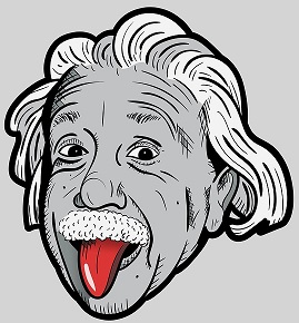
What if the popular persons had Alzheimer’s? What if Sir Arthur Conan Doyle had Alzheimer’s? The high functioning sociopath, Sherlock Holmes would be an unheard consulting detective. What if Einstein had Alzheimer’s and made the equation of the theory of special relativity? Imagine on a press meet one asks “Dear Einstein could you please explain the equation you deduced?” Oh! It’s simple E=…. E=… well I forgot. What a terrible situation. What if Stan Lee had Alzheimer’s? He might not be able to recognise his baby ‘Spidy’. Thank god, the only part worked super cool in Prof. Stephen Hawking was his brain after he was diagnosed with amyotrophic lateral sclerosis.
So what is Alzheimer’s???
Alzheimer’s disease is a type of dementia that causes problems with memory, thinking and behaviour. This neurodegenerative disease is characterized by two hallmark pathologies: β-amyloid plaque deposition and neurofibrillary tangles of hyperphosphorylated tau.
Aβ is formed from endoproteolysis of APP, a type 1 membrane protein. APP can be processed by a nonamyloidogenic pathway or an amyloidogenic pathway. In the non-amyloidogenic pathways, which is the most common, APP is cleaved by the α-secretase enzymes, which cut APP in the middle of the Aβ sequence, therefore preventing the formation of Aβ. In the non-amyloidogenic pathway APP is cleaved by BACE1, at the beginning of the Aβ sequence, thus liberating βAPP and a small carboxiterminal fragment, C99. Subsequently, C99 is further cleaved by the γ-secretase complex, generating Aβ40 and Aβ42. Aβ42 is the major species that accumulates in the AD brain. The major constituent of NFTs, the microtubule-associated protein tau, is hyperphosphorylated in the brains of AD patients. Tau protein normally stabilizes the microtubules in the neurons. This hyperphosphorylation is thought to contribute not only to the deregulation of microtubule dynamics, but also to tau polymerisation and aggregation.
What is doing out there???
Biomarkers and diagnostic tests are being developed to monitor the presence and progression of the disease. Positron emission tomography (PET) enables to image and measure amyloid and tau proteins in the brain of living patients. Cerebrospinal fluid is also used to measure other biomarkers. However, both tests are expensive.
Research is accelerating to develop a test just like the blood test to find out cholesterol and glucose level. To motivate such development ADDF (Alzheimer’s Drug Discovery Foundation) has teamed up with Bill Gates and other philanthropists. May be who knows Google may come up with the Artificial Intelligence just like the one which predicts heart disease by scanning our retina.
Latest studies!!!!!

Interesting study shows that aerobic exercises slightly reduced the amyloid buildup in the hippocampus- the part of the brain involved in memory. The reason is not clear yet.
Story
Let me tell you an interesting story of an Alzheimer’s researchers whose husband is a study subject.
This is the story of a 68-year-old neurology professor at Harvard Medical School, Dr. Dorene Rentz. She is a researcher involved in the A4 (Anti-Amyloid Treatment in Asymptomatic Alzheimer’s)study, a study that tests whether a drug called solanezumab can slow memory loss in people with elevated levels of amyloid plaque in their brains. Among the hundreds of participants undergoing the trail her 73-year-old husband, Ray Berggren is a part.

The participants get infusions of either solanezumab or a placebo. There are 1,169 participants, all of them 65 to 85 years old. The study is a double-blind, randomized control trial, so neither participants nor researchers know if patients receive the drug or a placebo.
Mr. Berggren is a retired auditor and database administrator. The first sign of exhibiting memory problems was when his son gave him a camera for Christmas four years ago. A couple of months later his son asked for the same camera and Mr. Berggren was unable to recollect the memory of the camera.
However, Mr. Berggren is now feeling good and handles grocery shopping, cooking at home and manages to function independently.
Let me remind you one more time that it’s an unsolved case and open for solving. Be like Sherlock Holmes!!!
Some topics in science always fascinate me and such a topic I read recently was about a transmembrane protein “The multi-drug transporters”. The first thing I do to make an acquaintance with any topic is going through some YouTube videos particularly through one of my favourite channel “Ben1994”. After that I go to Google scholar to find interesting research articles. The one I chose to read was “The role of multi-drug transporters in neurotherapeutics” published on 2009, which I felt like very good one.
Let me share some information about this awesome protein. It’s a glycoprotein found on the membrane of many cells. It’s a family of proteins.
The one type of protein family that we are familiar with is ABC (ATP Binding Cassette) transporters, the largest family of transmembrane proteins involved in the transport of molecules across membranes. The large ABC family has seven subfamilies ranging from ABCA to ABCG. Most but not all ABC transporters are multi-drug transporters. In human the most common multi-drug transporter is p-glycoprotein.
These glycoproteins are found on the apical portion of enterocytes (epithelial cells in intestine), the endothelial cells of blood capillaries in brain (blood brain barrier), hepatocytes lining the bile canalicus, the proximal convoluted tubule of nephrons etc.
Multi-drug transporters act like pumps that expel lipophilic/hydrophobic molecules out of the cell. It’s like a defence system to protect our cells from xenobiotics. It is this feature that makes it a boon and a curse.
In cancer and neurological disorders this transporter protein eliminates the drug from reaching the internal of the cell. Several inhibitors were created against this glycoprotein and the most recent being voruciclib (a phase 1 clinical trial drug normally inhibiting CDK4/6) which increased the intracellular accumulation and decreased the efflux of anticancer drugs. Still research is going on to find an efficient method to get rid of this wonderful transmembrane protein’s curse.
Why we use isopropanol for DNA precipitation? Why not ethanol? Since both are alcohol? Is this isopropanol so special? Let’s see.
Things we know.
What is electronegativity??
“Electronegativity means the tendency of an atom to attract the electrons towards itself in a chemical bond.”
Meaning – Oxygen atom pulls the electrons towards itself making it partially negative and hydrogen atom becomes partially positive. But the atoms do not become ionized.
The partially positive hydrogen atoms interact with the negatively charged phosphate groups of DNA making DNA soluble in water.
Inorder to precipitate the DNA we need to make the DNA insoluble in a solvent. For this we use alcohol.
Now arise the question ethanol or isopropanol?
In the typical precipitation step we add a salt. It may be sodium chloride, sodium acetate, lithium chloride, ammonium acetate.
In the case of using sodium acetate the sodium ions interact with the negatively charged phosphate groups of DNA.
If the medium there is water the force of attraction between sodium ions and phosphate groups of DNA will be very less because of the Coulomb’s law.
What is this law?
A law stating that like charges repel and opposite charges attract. The force of ionic interactions (Na+ and PO4-) in a solution depends on the magnitude of the charges, the distance between the charged groups, and the dielectric constant of the solvent in which the interaction occurs.

Coulomb’s constant is given by k= 1/4πεr
εr = relative permittivity
Relative permittivity is also commonly known as dielectric constant.
It is this dielectric constant that makes the difference.
Water has high dielectric constant, means that substances whose molecules contain ionic bonds (the salts used here) will tend to dissociate more in water yielding solution containing ions. Also the water will interact with these ions making them unavailable to neutralize the negative charge on DNA.
The dielectric constant of water is ~80, that of ethanol is ~24 and for isopropanol is ~18.
F = q1q2/r24πεwater | F = q1q2/r24πεethanol | F = q1q2/r24πεisopropanol
F = q1q2/r24π80 | F = q1q2/r24π24 | F = q1q2/r24π18
From these equations it comes to the conclusion that the force of attraction between sodium ions and phosphate groups of DNA is low compared to that in ethanol and isopropanol.
This is why isopropanol used instead of ethanol.
A biorepository is a laboratory facility to collect, process, store and distribute biospecimens such as urine, blood, tissues, cells, DNA, RNA, proteins from plants, animals, and human. These biospecimens and their related information (example: medical information in the case of human samples) are used for future research purposes.
“Biorepositories are libraries where biospecimens are stored and made available for clinical and research purposes.”- National Institutes of Health (NIH)
The primary functions of biorepositories are-
Biorepositories with only human specimens and data are referred to as biobanks.
With the advent of Next Generation Sequencing and high throughput molecular analysis, it is now possible to study the genomics of pathogens and particular disease conditions The genetic information generated helps to develop personalized medicines that why the biobanks have become an important resource for researches to answer many questions.
Tissue banks –
One specific type of biobank where biomedical tissues are stored under cryogenic conditions.
There are many ethical issues associated with biorepositories-
“There’s Plenty of Room at the Bottom” – Richard Feynman
Everyone knows about the light microscope which uses visible light and electron microscope which uses electron beams.
Keeping that as the base let’s discuss about what Electron tomography actually means.
The concept of matter waves by Louis de Broglie in 1924 led to the development of transmission electron microscopy (TEM) and the first TEM was invented by Ernst Ruska in the early 1930s. Since then many improvements have increased the resolution of TEM. In parallel with developments in TEM, scanning transmission electron microscopy (STEM) was introduced by Crewe et al.[1]
These inventions had opened new horizons to visualize objects at a grater resolution.
While TEM and STEM gives a nanometer scale length two dimensional images of the three dimensional samples, a third dimension is often required to determine the functionality of many structures.
It’s quite challenging to determine the atomic-resolution structure of bio-molecules with TEM because of the limitation in increasing the illumination dose.
‘The electron beam breaks the bond between the atoms in the structures.’
However in Electron tomography the exposure is applied at a lower dose rate over a longer time.
The term tomography means “imaging by sectioning”. The basic concepts of tomography were applied to TEM images for 3D analysis in the 1960s.
ET is similar to computerized tomography (CT). In CT the imaging equipment is moved around the patient to generate different images of a slice of the brain. However in ET the specimen is tilted incrementally in multiple directions with in the electron microscope to produce TEM images from different perspectives to reconstruct the object in its entirety (3D).[2]
The picture below shows the difference between images obtained through TEM and Electron tomography.

ET helps to generate detailed organellar characterization at the nanoscale and is essential for confirming direct membrane contacts between two organelles along the Z-axis with no loss of information due to sectioning.
The enormous technological advancements in instrumentation, computational power, and reconstruction algorithms have benefited ET in the past decades.
A wide variety of biological molecules, from a single macromolecule to a whole cell was studied effectively with ET. This technique has also enabled the study of conformational dynamics based on the structure of a single protein.
Software packages and algorithms developed for 3D reconstruction of objects. [1]
References
The three domain system introduced by Carl Woese et.al divides the cellular life forms into three domains – Archaea, Bacteria and Eukarya. Main difference is that the eukarya domain has the presence of membrane bound organelles. However this distinction was challenged after the discovery of a prokaryote phylum (planctomycetes) showing some features of eukaryotes.
The characteristics of planctomycetes have also found in other bacterial phyla and this has created a superphylum called PVC (Planctomycetes, Verrucomicrobiae and Chlamydiae) that include Planctomycetes, Verrucomicrobiae, Chlamydiae, Poribacteria, Lentisphaerae and the candidate phyla Omnitrophica.
The special features of these organisms differentiate them from the bacteria and make them more similar to archaea or eukaryotes.
A controversy has arised with the discovery of the PVC superphylum about the cell complexity emergence, allowed the occurence of the Cauldron hypothesis, which suggest continuity between three domains of life.
Hey there it’s me Vipin, a scholar who frequently search for new knowledge and ideas in Biology. The fictional character I like the most is Sherlock Holmes and I love to perform like him.
